Animal: Feline, male, DSH, 17 years old
History: Found dead with no medical history.
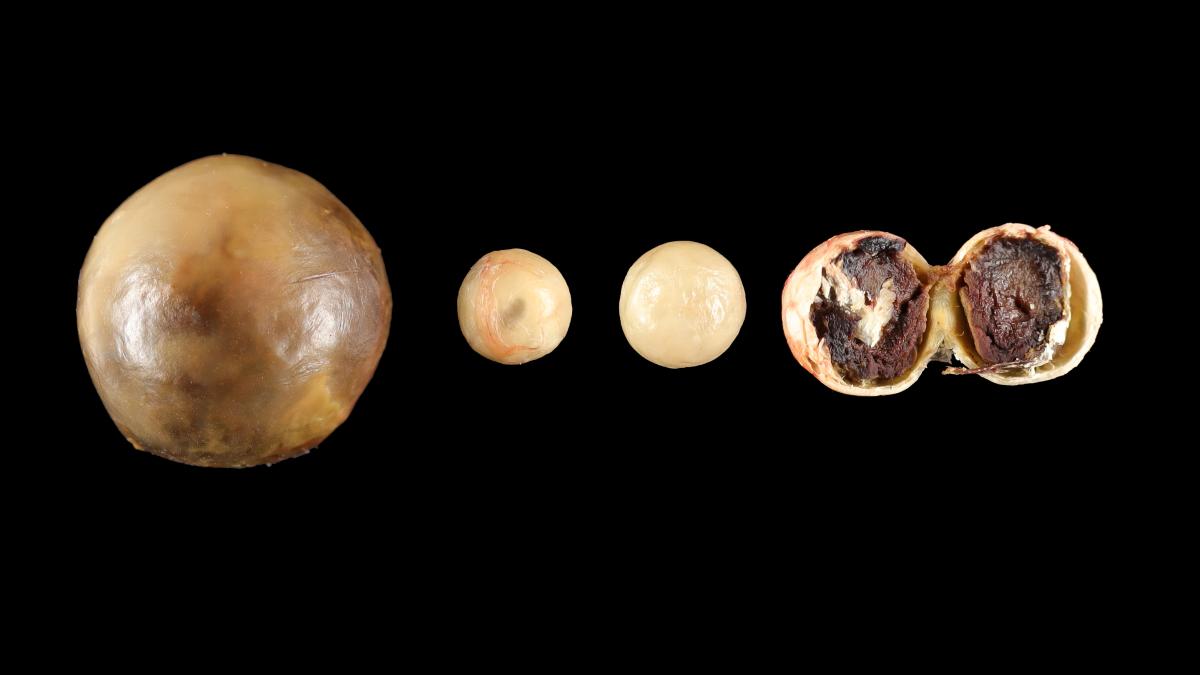
Macroscopical findings: The animal was in poor body condition and cachectic. Necropsy revealed a ruptured hepatic tumor associated with a hemoabdomen. Bilateral marked renal fibrosis and bilateral thyroid nodules were also found. In addition, four free smooth and spherical masses were present in the abdomen, ranging from 6 to 16 mm in diameter. They had an external solid and mineralized shell and a brown pasty and granular content (Bate's bodies).
Diagnosis: Multiple peritoneal Bate's bodies (nodular fat necrosis)
References:
SCHWARZ T., MORANDI F., GNUDI G., WISNER E., PATERSON C., SULLIVAN M., et al. Nodular fat necrosis in the feline and canine abdomen. Veterinary Radiology & Ultrasound. 2000, 41, 335 339.
SZABO D., FISCHETTI AJ. What Is Your Diagnosis? Peritoneopericardial diaphragmatic hernia and abdominal nodular fat necrosis (Bate’s bodies). Journal of the American Veterinary Medical Association. 2013, 244, 157 159.
Contributor: Edouard REYES-GOMEZ, Ecole Nationale Vétérinaire d'Alfort (Paris), FRANCE