
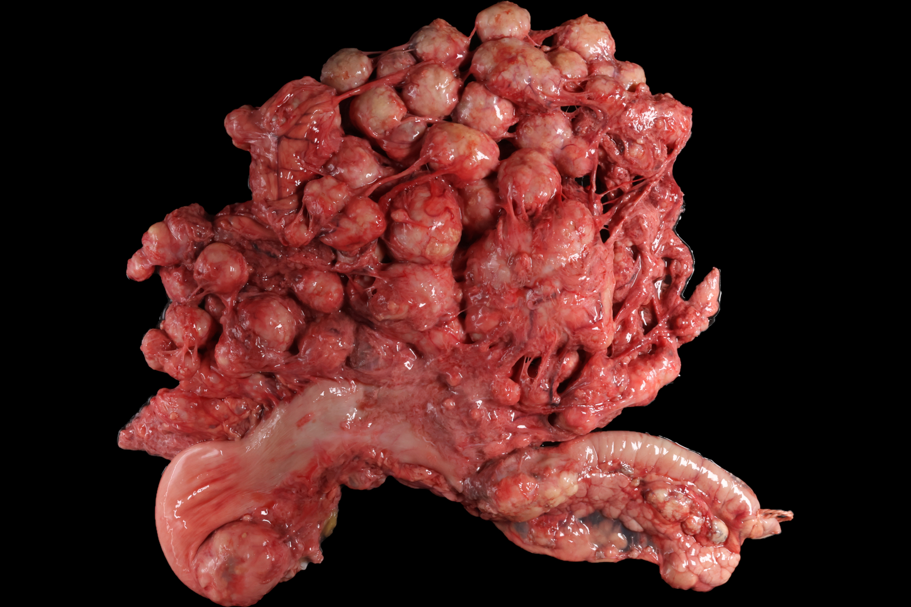
Animal: Dog (Canis lupus familiaris, mixed breed, female, 3 years old, 19 kg) Organ: Mesentery History: Animal was euthanized after sonography revealed a destructive process in the abdominal cavity Etiology: Larval stages of Echinococcus multilocularis
Etiologic diagnosis: peritoneal alveolar echinococcosis
Name of the disease: alveolar echinococcosis Macroscopic findings: 40-50 round, white to tan, 0.5 - 10 cm in diameter, firm, partially cystic but mostly solid nodules filled with scant to moderate amounts of clear to white, mucinous fluid
Morphologic diagnosis: Peritoneum: alveolar hydatid cysts, coalescing, chronic, severe with fibrosis and granulomatous inflammation
Comment: Within the normal life cycle of Echinococcus multilocularis canids such as foxes and dogs serve as definitive hosts and harbour the adult cestodes in the intestine. Usually without clinical signs, but with shedding of infectious and zoonotic eggs. Following accidental ingestion of these eggs, dogs may exceptionally act as aberrant intermediate and mostly dead-end hosts. The released oncospheres penetrate the intestinal wall and migrate to organs like liver and lung, where they develop into infiltratively growing metacestodes. The infiltratively growing metacestodic tissue is referred to as an alveolar hydatid (cyst), alveolar cyst, alveococcus, alveolar echinococcus or Echinococcus alveolaris and often mimics metastatic neoplasia.
Necropsy performed and picture prepared by: Dr. med. vet. Benjamin Diehl, Dipl. ECVP and Dr. Reiner Ulrich, Ph.D., Dipl. ECVP, Institute of Veterinary Pathology, Leipzig University
Bibliography: Corsini M, Geissbühler U, Howard J, Gottstein B, Spreng D, Frey CF. Clinical presentation, diagnosis, therapy and outcome of alveolar echinococcosis in dogs. Vet Rec. 2015 Dec 5;177(22):569. doi: 10.1136/vr.103470.