Signalment: Dog, female, adult, 8.9 kg
Diagnosis: Left adrenal gland: pheochromocytoma
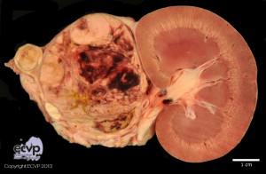
Description: Macroscopically the medulla of the left adrenal gland was replaced by a homogenous, soft, beige mass. Pararenal there was an expanding, encapsulated and well demarcated mass of up to 6.0 x 5.0 x 5.0 cm in dimension. Multifocally, on the cut surface hemorrhages and cystic areas were present. The right adrenal gland was of normal appearance.
Histologically the mass was highly cellular and composed of lobules of mostly round to polyhedral cells with palely eosinophilic, finely granular cytoplasm and round to ovoid hyperchromatic nuclei. Anisocytosis and anisocaryosis were moderate. The mass within the original adrenal gland showed the same features and was surrounded by a thin compressed rim of adrenal cortex. No invasions in other organs such as the left kidney or vessels were observed.
Comment: Pheochromocytomas are the most common neoplasm in the adrenal medulla of animals and develop most often in cattle and dogs. They are tumors of chromaffin cells and can be either unilateral or bilateral often with diameters of 10 cm or more. Affected dogs are usually middle aged with no breed or sex predilection. Functional tumors can secrete epinephrine and/or norepinephrine. Clinical signs attributed to catecholamine release are rare. 39 to 50% of the canine tumors invade the Vena cava, liver or kidney by local extension. Invasion and thrombosis of the Vena cava or extramural compression can occlude venous return from posterior extremities, causing edema.
Image & Authored by: Stefanie Binder, Department of Veterinary Pathology, Freie Universität Berlin, Germany