Animal: Canine, 6 years old
Organ: Small intestine
History: Vomiting, anorexia, multiple intestinal masses, resection of the pathological segments
Macroscopical findings: 2 jejunal segments of 35 cm respectively 7 cm in length, with 5 masses in total, measuring between 2 and 7 cm in diameter. Cross sections of the masses showed massive focal extensive thickening of the intestinal wall, firm, tan, with narrowing of the intestinal lumen. The masses had been completely excised. One biopsy of mesenteric lymph node, no lesions noticed.
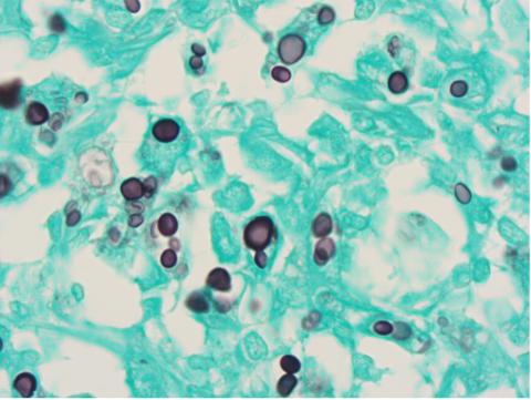
Histopathological findings: Masses composed of nodular infiltrates of mainly macrophages, lesser numbers of lymphocytes, plasma cells and neutrophils, with granulation tissue. In the cytoplasm of macrophages or free between cells, there are numerous mycotic agents, they are stainless spores, sometimes showing narrow based budding. Size of the spores varies between 3 and 15 micrometre. They are sometimes surrounded by a thick optically empty capsule. The spores are PAS and Grocott positive and their thick capsule is positive for mucicarmine.
Lymph node reactive.
Diagnosis: Multifocal, severe, chronic granulomatous enteritis with numerous intralesional spores of mycotic agents showing narrow based budding, morphology, and staining properties consistent with Enteric Cryptococcosis (C. Neoformans)
Contributor: Nicolas Kühn and Maria Wegelin, DVM, Dipl. ECVP, Kühn Pathologie AG laboratory, Hünenberg, Switzerland