
Animal: Cat, intact male, no breed, 13 years old
A scrotal tissue fragment measuring 3.1 × 2.2 × 1.3 cm was submitted for histopathological examination. The clinical history indicated recurrent cystic lesions of the scrotum. The testes were not included in the specimen.
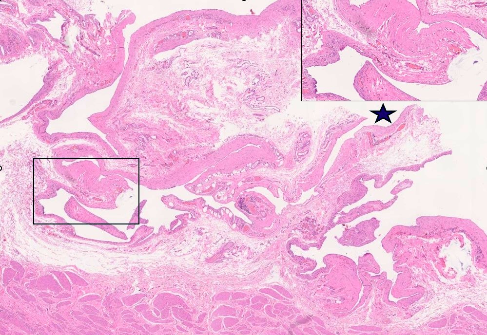
Within the mid-depth of the scrotal wall, a markedly anfractuous cavity was identified, without evident luminal content. The cavity wall was predominantly composed of moderately dense connective tissue and was lined by endothelial/mesothelial-type cells (see inset). Polypoid projections into the lumen were observed. The communication with the scrotal sac is presumed to be located in the area marked with a star. The remaining inner surface of the scrotum, on both sides of the cavity opening, formed deep indentations. No neoplastic cells were identified.
Diagnosis:
Intrascrotal cystic invagination of the external leaflet of the tunica vaginalis, with secondary distension possibly due to hydrocele.
The findings described above appear to represent a congenital malformation, for which no specific references were identified in the literature. We believe that this anomaly had not been previously detected, possibly because there had been no noticeable change in scrotal size or contour. Its recent detection may have been precipitated by fluid accumulation between the layers of the tunica vaginalis (hydrocele), either of local or peritoneal origin, which likely filled the space created by the invagination. In the present case, the origin of the hydrocele remains undetermined. Surgical excision of the lesion is considered curative.
Authorship (text and photos) – Maria C. Peleteiro and Sofia Valente
URANOLABPT, Avenida Pedro Álvares Cabral, Centro Empresarial Sintra Estoril VI V E23, 2710-297 Sintra
Contact - mcpeleteiro@gmail.com